
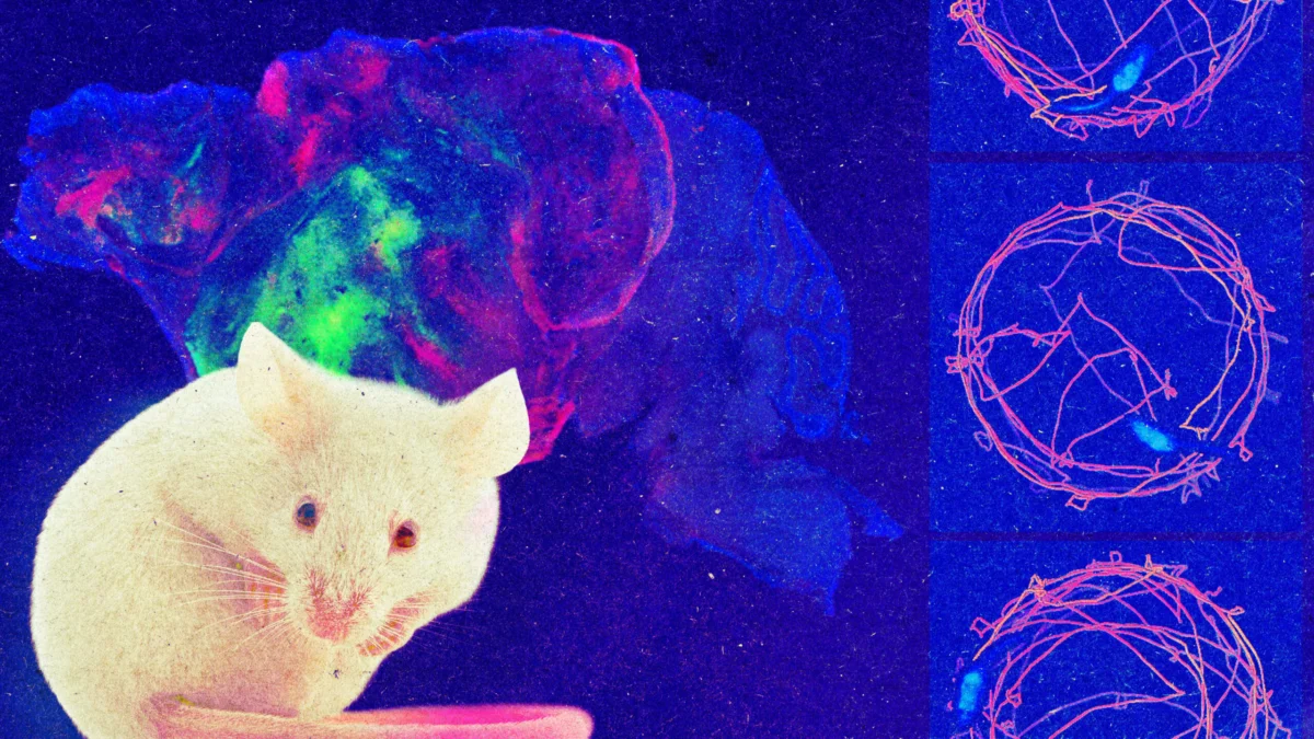
In a landmark study published in the prestigious journal Nature, a research team at Stanford University has unveiled a significant leap forward in the field of neuroscience and interspecies chimera research. Led by prominent neuroscientist Dr. Sergiu Pașca, the laboratory has successfully integrated human neural tissue into genetically engineered mice that were selectively bred to lack key structures in their native brains. This latest achievement moves beyond the team’s previous milestones of introducing human brain "organoids"—three-dimensional, miniaturized clusters of lab-grown neural tissue—into the brains of healthy infant rodents. By deliberately creating neurodevelopmental voids within the host animals, the researchers have allowed human cells to proliferate, colonize, and functionally integrate on an unprecedented scale.
The publication of these findings has immediately reignited global scientific and bioethical debates regarding the boundaries of regenerative medicine, the manipulation of species barriers, and the philosophical implications of blending human and animal neural architecture. While proponents highlight the immense value of these "xenocortical mice" for modeling complex human psychiatric and neurological disorders, ethicists and researchers alike are grappling with the fast-evolving capabilities of modern biotechnology and the profound questions they raise about consciousness, identity, and the moral status of modified organisms.
Chronology of Human-Rodent Neural Chimerism
The foundation for this breakthrough was laid over several years of rigorous experimentation by Dr. Pașca and his colleagues at Stanford University. The timeline of this research reflects an accelerating trajectory in stem cell biology and neuroengineering:
- Initial Organoid Development: Years prior to the current study, Dr. Pașca’s laboratory pioneered methods to derive complex three-dimensional neural tissues from human induced pluripotent stem cells (iPSCs). These organoids successfully mimicked various regions of the human brain, including the cerebral cortex, exhibiting spontaneous electrical activity and cellular stratification.
- Early Transplantation Milestones: In a widely publicized study, the Stanford team demonstrated that human cortical organoids could be successfully transplanted into the brains of newborn rats. Over several months, these human cells survived, vascularized, and established functional neural connections with the host rodent brain circuitry, even influencing the animals’ behavioral responses to sensory stimuli.
- Genetic Modification of the Host: Seeking to overcome the physical and spatial limitations of injecting human tissue into an already fully populated rodent brain, the researchers engineered a novel mouse model. These mice were genetically modified so that critical cells within the cortex and hippocampus—the regions governing sensory processing, higher cognition, and memory—failed to develop properly.
- Publication in Nature: The culmination of these efforts was formally reported in Nature, detailing how human cells transplanted into these structurally deficient brains expanded to occupy the vast majority of the vacant neurological space, fundamentally altering the functional capacity of the host animals.
Methodological Breakthroughs and Experimental Findings
The core innovation of the recent study lies in the deliberate creation of a structural niche. In standard neurobiology, introducing foreign tissue into an intact mammalian brain meets fierce competition from resident cells, limiting the integration and growth of the transplant. By utilizing genetically modified mice missing the vast majority of cells in both the cortex and the hippocampus, Dr. Pașca’s team engineered an environment highly receptive to foreign colonization.
When human neural organoids were introduced into these specially prepared hosts, the response was dramatic. Rather than being rejected or remaining dormant, the human cells actively divided, grew, and migrated throughout the empty cavities. Within weeks to a few months, human neural tissue had taken over the majority of the space traditionally reserved for the mouse cortex and hippocampus.
Behavioral observations of the host animals yielded fascinating and nuanced results. Mice lacking cortical and hippocampal tissue naturally exhibit severe cognitive deficits. In baseline evaluations, such as maze navigation tests, these modified mice demonstrated profound memory impairments, unable to retain spatial awareness of areas they had previously explored. Remarkably, when these same structurally deficient mice were implanted with human neural organoids, their performance on maze tests improved significantly. This behavioral recovery provided concrete empirical evidence that the human tissue was not merely surviving inertly, but was actively participating in and rescuing the cognitive functions of the host animal.
Despite these remarkable changes, the mice retained their fundamental rodent behaviors. They moved about their enclosures and vocalized normally, showing no outward signs of human-like behavior or consciousness. Dr. Pașca attributes this to the vast evolutionary distance between humans and mice, as well as the constrained physical scale of the rodent cranium, which limits the total volume of human tissue that can be supported.
Broader Context and Technological Implications
The creation of xenocortical mice occurs against a backdrop of rapid convergence between biotechnology, artificial intelligence, and neuroscience. Labs around the world are increasingly pushing the limits of what lab-grown human brain tissue can achieve. Recent experiments have explored connecting brain organoids to silicon chips to process digital information, play video games, or decode speech patterns. Concurrently, medical researchers have proposed utilizing neural organoids as biological replacement parts to repair damage caused by strokes, traumatic brain injuries, or neurodegenerative conditions such as Alzheimer’s and Parkinson’s disease.
Carsten Charlesworth, a Stanford researcher not involved in the study, points out that the sheer adaptability of human neural tissue across a species barrier is the most striking takeaway from the research. The ability of human cells to cross the mammalian developmental divide, wire into a foreign nervous system, and restore lost function underscores the sheer potency of modern genetic engineering and stem cell technology. As these tools become more refined, they inevitably challenge traditional scientific and philosophical assumptions regarding the uniqueness of human neural circuits and the definitions of biological identity.
Furthermore, the potential applications for studying human-specific neurological and psychiatric disorders are profound. Many conditions, ranging from schizophrenia to severe autism, affect neural pathways that are uniquely developed in humans and difficult to model accurately in standard animal models. Xenocortical systems offer a living laboratory where human-specific cellular pathologies can be studied in an intact, functioning organismic context.
Ethical Frameworks and the "Red Line" on Primates
Recognizing the profound ethical implications of their work, Dr. Pașca and his peers have actively engaged with bioethicists to establish guardrails for the field. Last year, Dr. Pașca convened a specialized group of ethics experts to examine the horizon of neural organoid technology. Among the central topics discussed were the theoretical risks of an animal developing elements of human consciousness, as well as the commercial dangers of unregulated "organoid therapy clinics" potentially offering unproven, scam treatments to vulnerable and desperate patients.
At present, the researchers maintain that there is no credible risk that the rodents in these experiments possess human cognitive capacities or subjective experience. The tiny volume of the mouse brain, combined with the profound evolutionary gap separating rodents from humans, acts as a natural safeguard against the emergence of human-like sentience.
However, this exact biological calculation forms the basis for a strict ethical boundary concerning other species. Dr. Pașca has issued explicit warnings against scaling these experiments upward, specifically cautioning against the introduction of human brain organoids into primates, such as monkeys. Because non-human primates share a much closer evolutionary lineage, brain architecture, and physiological complexity with humans, the introduction of large volumes of functioning human neural tissue could potentially blur the cognitive and moral boundaries between human and animal subjects.
"One of the things that I see as a very clear red line is doing this experiment in a primate," Dr. Pașca stated regarding the future of the research. "I don’t think that is justified at this point in any way."
Conclusion and Future Outlook
The publication of the Stanford team’s findings in Nature marks a watershed moment in experimental neuroscience. By successfully merging human neural tissue with genetically tailored mammalian hosts, the research opens new avenues for therapeutic discovery while simultaneously testing the limits of contemporary bioethics. As the scientific community digests these results, the imperative to balance rigorous innovation with thoughtful oversight will only intensify. Researchers, ethicists, and regulatory bodies must navigate this uncharted territory carefully, ensuring that the remarkable power to reshape biology is guided by clear principles that respect the dignity of both human and animal life.